Stephen DiNardo, Ph.D.
1111 BRB II/III
421 Curie BoulevardPhiladelphia, PA 19104-6058
Office: use email
Fax: 215-898-9871
Research Area
Professor of Cell and Developmental Biology
Additional Appointment Titles
- Member, Penn Institute for Regenerative Medicine (IRM)
Graduate Group Affiliations
Education
- B.A. (Biochemistry)
Columbia University, 1977. - Ph.D. (Biochemistry/Molecular Biology)
Dept. of Biochemistry, SUNY at Stony Brook, Laboratory of Rolf Sternglanz, 1983. - Cert. (Unconscious Bias for Leaders – Impact on Decision-Making)
Perelman School of Medicine, 2020. - Cert. (iCARE Training on Crisis Management)
CAPS, Perelman School of Medicine, 2021. - Cert. (Mentoring Facilitator for Faculty)
CIMER, Univ Wisconsin, 2022.
Post-Graduate Training
- Helen Hay Whitney Postdoctoral Fellow, Dept. of Biochemistry & Biophysics, Laboratory of Patrick H. O’Farrell, UCSF, 1984-1986.
- Lucille P. Markey Scholar, Dept. of Biochemistry & Biophysics, Laboratory of Patrick H. O’Farrell, UCSF, 1986-1988.
Description of Research Expertise
Research Interests
- Niche Assembly & Stem Cell Control
Key words: Stem Cells, Niche, Morphogenesis, Cell Polarity
Description of Research
Niche – Stem Cell Interactions
Stem cells hold the key to the continual renewal of many of our tissues. In addition, the manipulation of these cells holds much promise for regenerative medicine. Unfortunately, there are severe limitations in our knowledge of the regulation of stem cells and that compromises our understanding of normal tissue renewal, and restrains their clinical utility. To uncover the general principles of stem cell regulation, we study adult stem cells within their natural environment, the niche, by using Drosophila spermatogenesis as a model system. This allows us to apply both genetic and genome-scale molecular approaches to uncover novel factors governing niche-stem cell interactions, self-renewal, differentiation, proliferation and stem cell aging. Most importantly, it allows for high-resolution, real-time imaging of the initial assembly of the niche, and of stem cell behavior. Given the deep conservation of mechanisms across species, we are confident that concepts revealed by our studies will apply to mammalian stem cell – niche interactions.


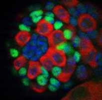
Among the strengths of this system are the molecular markers and techniques to unambiguously identify the niche cells, as well the two stem cell lineages that maintain this tissue. The micrograph shows the tip of a testis (left) where the central cluster of cells (blue-green) comprise the niche (called a “hub”); the germline stem cells (GSCs) map to the first tier of (red) cells surrounding the niche, and these GSCs are intermingled with somatic stem cells (green), which express the somatic stem cell-specific factor Zfh-1. Another advantage to studying this system is that we can culture the testis and live-image the production of new stem cells (come see some gorgeous movies made by Gabby https://biorxiv.org/cgi/content/short/2024.06.04.597489v1). And, excitingly, we have access to a testis single cell RNA sequence data set to help identify new regulators!
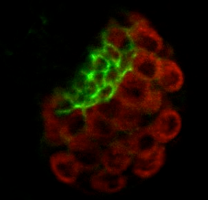
Given that the niche is such a critical component regulating stem cell behavior, we also study how its cells are first specified, and how they assemble to form a functional niche. The right-hand micrograph shows a gonad with newly specified niche cells, marked by a cell surface protein (green). Again, combining genetics with lineage tracing and live-imaging we have uncovered several circuits necessary to specify these cells, and are studying how the individual cells migrate directionally and coalesce together to form the niche (come see some gorgeous movies made by Bailey!
(https://biorxiv.org/cgi/content/short/2023.09.08.556877v2). We have recently learned that that coalescence involves cell biological and cell mechanical interactions between niche and stem cells.
Finally, we are extending our studies to the assembly of the insect hematopoietic niche. This niche controls the progenitors for the innate immune system, and is comprised of a bilaterally-symmetric set of cells specified in late-stage embryos. Come see some gorgeous movies made by Kara showing how these cells migrate and coalesce to take up their final positions in the lymphatic organ. Kara is exploring how extracellular signals govern the precise positioning of this niche (https://biorxiv.org/cgi/content/short/2024.06.21.600069v1).
Lab Personnel
- PhD Student
- Bailey Warder
- Gabriela Vida
- Kara Nelson
- Research Assistant
- Chung-Tsai Lee (Lee)
Don’t forget Steve, he’s at the bench, too!
Limited Rotation Projects for 2024-2025; see Steve
- Niche – Stem Cell interaction projects:
Students will use genetic and genome-scale molecular techniques, combined with live-imaging, to identify factors 1) that first specify Niche cells, 2) that organize those cells into a functioning niche, and 3) that regulate the balance between self-renewal and differentiation within the stem cells. Students can dive into functional studies already ongoing in these areas. Come by to see Lauren & Bailey’s neat movies of a niche first being formed, as well as Gabby and Liz’s beautiful movies of action at a stem cell niche!! As work progresses, we envision collaborative opportunities for work, where students can explore how the concepts we are revealing in this Drosophila system apply to other niche-stem cell systems investigated here at Penn. - Morphogenesis of a niche projects:
Students will ask how signaling pathways control the shape and behaviors of niche cells in the animal, rather than in the contrived environment of a cultured cell. They will investigate how cell biological responses are coordinated between niche and associated stem cell. This involves examining the cytoskeletal changes that occur during cell rearrangement in real-time, using live imaging with Spinning Disk Confocal Microscopy (come by and see some of Bailey and Kara’s cool movies!). That approach is complemented by in vivo manipulation of target proteins of the key signaling pathways involved – proteins such as small GTPases, polarity proteins or adhesion and cytoskeletal components. In turn, those perturbations are coupled with high resolution imaging techniques to examine the changes at cell-cell interfaces, such as changes to cell bond tension by laser cutting or by FRET/FRAP, to map out how selective alterations of cyto-architecture affect tissue dynamics.
Selected Publications
Anllo L, DiNardo S.: Visceral mesoderm signaling regulates assembly position and function of the Drosophila testis niche. Dev Cell 57: 1009-1023, Apr 2022.
Raz AA, Vida GS, Stern SR, Mahadevaraju S, Fingerhut JM, Viveiros JM, Pal S, Grey JR, Grace MR, Berry CW, Li H, Janssens J, Saelens W, Shao Z, Hu C, Yamashita YM, Przytycka T, Oliver B, Brill JA, Krause H, Matunis EL, White-Cooper H, DiNardo S, Fuller MT.: Emergent dynamics of adult stem cell lineages from single nucleus and single cell RNA-Seq of Drosophila testes. Elife 12: e82201, Feb 2023.
Lenhart Kari F, Capozzoli Benjamin, Warrick Gwen S D, DiNardo Stephen: Diminished Jak/STAT Signaling Causes Early-Onset Aging Defects in Stem Cell Cytokinesis. Current Biology : CB 29(2): 256-267.e3, Jan 2019.
Kari F. Lenhart Stephen DiNardo: Somatic cell encystment promotes abscission in germline stem cells following a regulated block in cytokinesis. Developmental Cell 34(2): 192-205, July 2015.
*Anllo Lauren, *Plasschaert Lindsey W, *Sui Justin, DiNardo Stephen: Live imaging reveals hub cell assembly and compaction dynamics during morphogenesis of the Drosophila testis niche. Developmental Biology 446(1): 102-118, 02 2019 Notes: * Co-First Authors.
Ong Katy, Collier Camille, DiNardo Stephen: Multiple feedback mechanisms fine-tune Rho signaling to regulate morphogenetic outcomes. Journal of Cell Science 132(8), Apr 2019.
Wingert L; DiNardo S. : Traffic jam functions in a branched pathway from Notch activation to niche cell fate. Development. Company of Biologists, 142(13): 2268-77, July 2015.
Ly Dan, Resch Erin, Ordiway George, DiNardo Stephen: Asymmetrically deployed actomyosin-based contractility generates a boundary between developing leg segments in Drosophila. Developmental biology 429(1): 165-176, 09 2017.
Donoughe, Seth. DiNardo, Stephen.: dachsous and frizzled contribute separately to planar polarity in the Drosophila ventral epidermis. Development 138(13): 2751-9, Jul 2011 Notes: Faculty of 1000 manuscript.
Leatherman, J. L., DiNardo, S.: Germline self-renewal requires cyst stem cells and stat regulates niche adhesion in Drosophila testes. Nat Cell Biol 12: 806–811, August 2010 Notes: COVER photo.
Leatherman, J. L., DiNardo, S.: Zfh-1 controls somatic stem cell self-renewal in the Drosophila testis and nonautonomously influences germline stem cell self-renewal. Cell Stem Cell 3(1): 44-54, July 2008.

